
Page 60 - Micro5 Brochure 2017
P. 60
Effect of microcurrent electrical stimulation on tendon healing 115
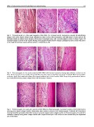
Fig. 4 Photomicrograph of a three week neotendon (H&E 200·). (A) Untreated control showing less-organized fibroploriferative
changes with poorly aligned collagen bands, inflammatory tissue reaction with mononuclear cells infiltrations is clearly noticed. (B)
Photomicrograph of cathodal group showing well-developed granulation tissue with a properly aligned pattern of collagen bands. (C)
Photomicrograph of anodal treated tendons showing well-organized fibroploriferative changes. Inflammatory tissue reaction with notice
of the newly formed blood vessels and few numbers of inflammatory cells.
Fig. 5 Photomicrograph of a five weeks neotendon (H&E 200·). (A) Untreated neotendon showing high cellularity in relation to the
fibers. Notice attempts to form bundles with parallel fibers but still in disarray. (B) Photomicrograph of cathodal MES showing cellular
neotendon, small blood vessels and collagen fibers appears scattered and in loose bundles. Notice foreign body granulomatous reaction.
(C) Anodal MES showing mature collagen fibers with fibrocystes in-between.
Fig. 6 Photomicrograph of an eight week neotendon (H&E 200·). (A) Photomicrograph of untreated tenotomized left Achilles tendon
showing poorly aligned collagen bundles. Inflammatory tissue reaction is observed. (B) Photomicrograph of cathodal MES stimulation
showing diminished granulation tissue with formation of properly aligned mature collagen bundles. (C) Photomicrograph of Anodal MES
stimulation showing closely packed collagen bundles with compressed fibrocytes. Both of them are well oriented along the longitudinal
axis of the tendon.